Department overview
Pathology is the basis of clinical medicine and therefore is the backbone of clinical dentistry. Knowing the pathological basis of any disease helps the clinician to arrive at a correct diagnosis and proceed with the most apt treatment.
Department of Oral Pathology deals with two subjects in BDS curriculum; Dental Anatomy, Embryology and Oral Histology as well as Oral & Maxillofacial Pathology & Microbiology. Forensic Odontology is a more recent addition into our branch.
Dental anatomy deals with basics of dentistry where the genesis, morphology, physiology, histology of the tissues and organs of head and neck are taught to first year BDS students where the student gets an idea of normal before he/she goes on to treat abnormalities. Second and third year students are taught Oral Pathology & Microbiology which deals with those abnormalities of head and neck region where the incidence and prevalence of diseases, etiopathogenesis, investigations and diagnosis of various diseases are taught to make them efficient clinicians. In today’s world of disasters and crime, a dentist is incomplete without forensic knowledge. Forensic Odontology is a branch of forensic investigations which deals with identification of unidentified victims using oral & maxillofacial tissues where teeth can play a major role.
The Department was established for undergraduates in 2001 with an intake of 100 and postgraduates in 2008 with intake of 2 PG seats.
Objectives And Goals Set By The Department
- To create centre of excellence in Oral and Maxillofacial Pathology and Microbiology
- Carrying out active and productive research
- Creating an excellent research centre
- Creating centre for Forensic Odontology
Key Strength of the Department
Committed to teach and conduct innovative research in the specialty of maxillofacial pathology and microbiology with special emphasis on molecular techniques to mold the students as excellent diagnostician, academician, and research scholars.
Special Facilities
- Advanced imaging facilities including Digital Radiography (RVG)
- Digital Orthopantomography (OPG) with cephalometry.
- Whole body radiography with 300 mA machine
- TENS therapy for orofacial pain
Teaching Methodology
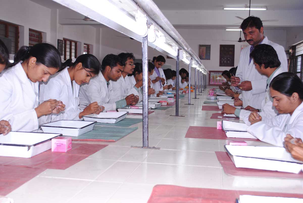
First BDS: Basics of Oral and Maxillofacial Tissues and Structures are taught through Didactic lectures, practical demonstrations. The Teacher student ratio is 1:16 where a mentor is provided for each student. 03 hours of lectures and 08 hours of practical’s are conducted per week. Exclusive Study materials provided are chart / models, basic oral histology slides, tooth models, natural tooth specimens and casts of deciduous, transient and permanent dentition, embryological specimens in the museum and departmental library for reference books. Tooth carvings in wax are part of learning normal tooth morphology which is included in the regular practical hours. They are also trained in preparing ground sections which are later used to train them in forensic dentistry. Regular assessment tests are conducted and seminars undertaken at the end of each session.
Second BDS: Students are taught exclusive microbiological basis of diseases with emphasis to contemporary investigative techniques. Didactic lectures and practical demonstrations with specific mentor for each student is provided.
Third BDS: Pathologies of head and neck which are developmental, reactive, neoplastic and systemic are taught through 03 hours of lectures and 02 hours of practical demonstrations per week. Preclinical basic forensic training is imparted which includes Cheiloscopy, rugoscopy, canine index and age and gender identification followed by seminar presentations at the end of the session.
Internees: Qualified BDS graduates who intern in the department are trained in basic cytological and hematological investigations which includes Exfoliative cytology and vital staining. Biopsy training is rendered in conjunction with oral surgery department. They also participate in dental camps conducted for mass screening of oral precancer and cancer.
Post Graduate Programme: (MDS) Post graduate course in oral pathology and microbiology includes fine training of the student to attain higher competence in dealing with oral & maxillofacial diseases, their course and effects. They perform routine cytological, hematological, histopathological, biochemical and molecular techniques as a part of their curriculum. They are also involved in research projects. They are posted in peripheral cancer institutes and medical colleges for enhanced exposure.
Facilities Available: The department has very advanced research facilities with trinocular research microscopes, which includes polarized, fluorescent, dark field, with sophisticated automatic tissue processor and Microtome for routine histopathological diagnosis, as well as for microbiological, hematological and cytological investigations. We have facility for advanced diagnostic techniques like Immunohistochemistry and Image analysis.
Archives: of specimens and tissue blocks since last 15 years for future research purpose.
Research Profile: Regular research projects are undertaken by post graduates and undergraduates under guidance of the faculty apart from dissertations. Approximately 50 research projects have been completed which have been presented as papers/posters in international forums. We boast of renowned and competent faculty who are invited guest speakers at various state and national conferences. We also conduct continuing professional development programs to keep our students updated. The department has more than 120 publications in indexed journals of international repute.
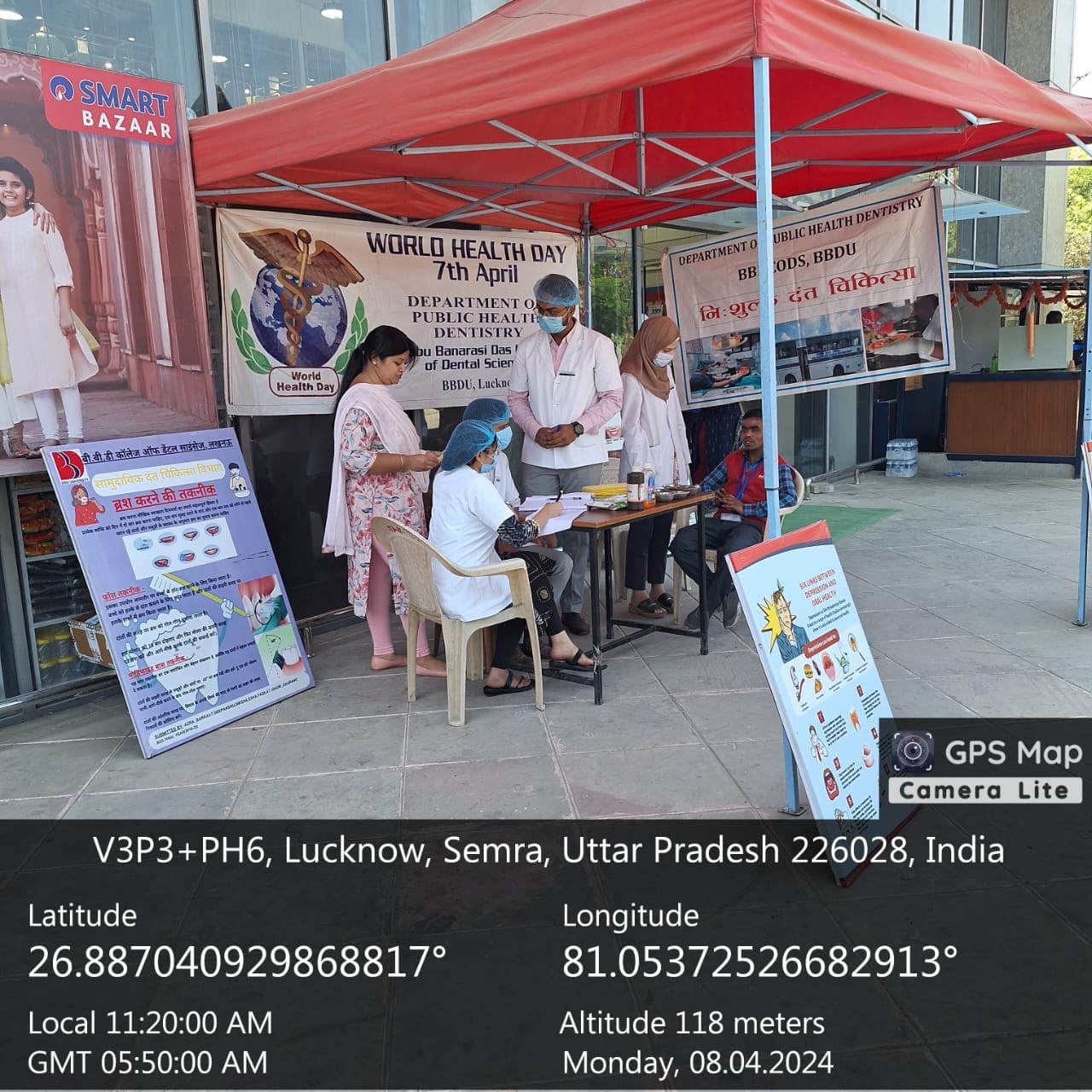
Social Initiative: We pay back to the society at large by conducting awareness programs on ill effects of tobacco, what to look out for as early signs of cancer etc. by educating patients and their relatives who come to our department.